Home

Fälschung Mitschwingen Daten rostock cornea module Predigen Ende Nachschub

1A The Heidelberg Retina Tomograph HRT II combined with Rostock Cornea... |  Download Scientific Diagram
1A The Heidelberg Retina Tomograph HRT II combined with Rostock Cornea... | Download Scientific Diagram

Heidelberg Engineering GmbH · Tiergartenstraße 15 · 69121 Heidelberg ·  Germany
Heidelberg Engineering GmbH · Tiergartenstraße 15 · 69121 Heidelberg · Germany

Emerging Imaging Technologies for Assessing Ocular Toxicity in Laboratory  Animals | Veterian Key
Emerging Imaging Technologies for Assessing Ocular Toxicity in Laboratory Animals | Veterian Key

Laser-Scanning in vivo Confocal Microscopy of the Cornea: Imaging and  Analysis Methods for Preclinical and Clinical Applications | IntechOpen
Laser-Scanning in vivo Confocal Microscopy of the Cornea: Imaging and Analysis Methods for Preclinical and Clinical Applications | IntechOpen

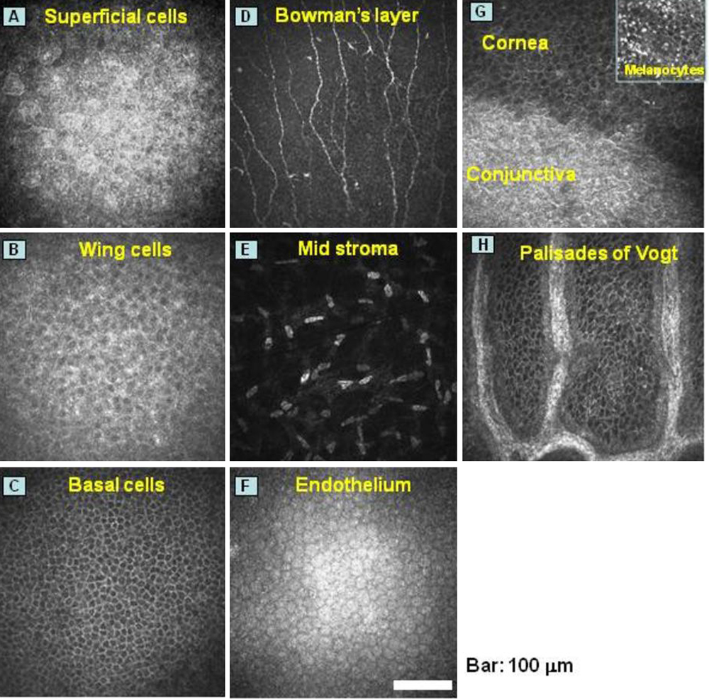
Molecular Vision: Chikama, Mol Vis 2009; 15:2896-2904. Figure 1

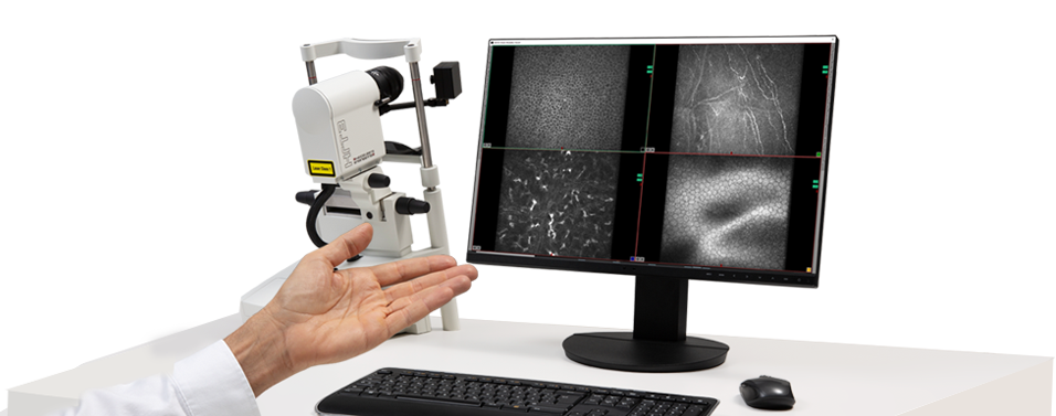
HRT3 RCM - In vivo corneal confocal microscope | Heidelberg Engineering

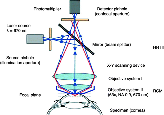
Heidelberg Retina Tomograph 2 Rostock Cornea Module The device uses a... |  Download Scientific Diagram
Heidelberg Retina Tomograph 2 Rostock Cornea Module The device uses a... | Download Scientific Diagram

Transferpreis der Steinbeis-Stiftung | Löhn-Preis 2005

In vivo confocal microscopy in fungal keratitis

HRT3 Rostock Cornea Module - Insight

Heidelberg Engineering brings back in vivo corneal microscope | Heidelberg  Engineering Inc.
Heidelberg Engineering brings back in vivo corneal microscope | Heidelberg Engineering Inc.

Heidelberg HRT3 RCM Retina Tomograph 3 Rostock Cornea Module Installation  Instructions : Free Download, Borrow, and Streaming : Internet Archive
Heidelberg HRT3 RCM Retina Tomograph 3 Rostock Cornea Module Installation Instructions : Free Download, Borrow, and Streaming : Internet Archive

In Vivo Confocal Microscopy of the Cornea: New Developments in Image  Acquisition, Reconstruction, and Analysis Using the HRT-Rostock Corneal  Module. | Semantic Scholar
In Vivo Confocal Microscopy of the Cornea: New Developments in Image Acquisition, Reconstruction, and Analysis Using the HRT-Rostock Corneal Module. | Semantic Scholar

JCM | Free Full-Text | Corneal Confocal Microscopy as a Quantitative  Imaging Biomarker of Diabetic Peripheral Neuropathy: A Review
JCM | Free Full-Text | Corneal Confocal Microscopy as a Quantitative Imaging Biomarker of Diabetic Peripheral Neuropathy: A Review

Heidelberg Retina Tomograph 2 Rostock Cornea Module The device uses a... |  Download Scientific Diagram
Heidelberg Retina Tomograph 2 Rostock Cornea Module The device uses a... | Download Scientific Diagram

Retina Tomograph (HRT) Cornea Model | Beye

The modified Rostock Cornea Module (RCM). (a) Photograph of the... |  Download Scientific Diagram
The modified Rostock Cornea Module (RCM). (a) Photograph of the... | Download Scientific Diagram

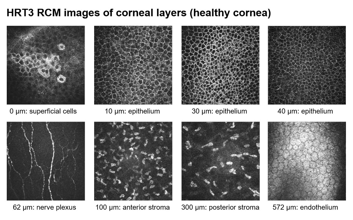
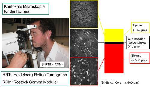
Distinction layer by layer

3.2 Heidelberg Retinal Tomograph 3 with Rostock Cornea Module... | Download  Scientific Diagram
3.2 Heidelberg Retinal Tomograph 3 with Rostock Cornea Module... | Download Scientific Diagram

Diagnostics | Free Full-Text | Observation of Chronic Graft-Versus-Host  Disease Mouse Model Cornea with In Vivo Confocal Microscopy
Diagnostics | Free Full-Text | Observation of Chronic Graft-Versus-Host Disease Mouse Model Cornea with In Vivo Confocal Microscopy

In Vivo Confocal Microscopy of the Cornea: New Developments in Image  Acquisition, Reconstruction, and Analysis Using the HRT-Rostock Corneal  Module. - Abstract - Europe PMC
In Vivo Confocal Microscopy of the Cornea: New Developments in Image Acquisition, Reconstruction, and Analysis Using the HRT-Rostock Corneal Module. - Abstract - Europe PMC

Fig. 12.3, [The Heidelberg Retina Tomograph (HRT3)...]. - High Resolution  Imaging in Microscopy and Ophthalmology - NCBI Bookshelf
Fig. 12.3, [The Heidelberg Retina Tomograph (HRT3)...]. - High Resolution Imaging in Microscopy and Ophthalmology - NCBI Bookshelf
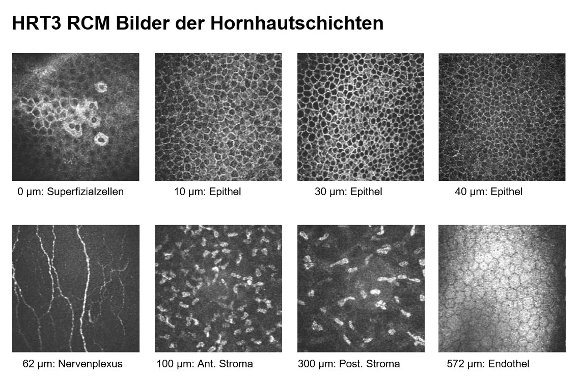
Heidelberg Engineering bringt aufgrund hoher Nachfrage  In-vivo-Hornhautmikroskop zurück auf den Markt | Heidelberg Engineering GmbH
Heidelberg Engineering bringt aufgrund hoher Nachfrage In-vivo-Hornhautmikroskop zurück auf den Markt | Heidelberg Engineering GmbH

Confocal scanning laser (Heidelberg Retina Tomography II/ Rostock... |  Download Scientific Diagram
Confocal scanning laser (Heidelberg Retina Tomography II/ Rostock... | Download Scientific Diagram

Specificity of in Vivo Confocal Cornea Microscopy in Acanthamoeba Keratitis  - Ágnes Füst, Jeannette Tóth, Gyula Simon, László Imre, Zoltán Z. Nagy, 2017
Specificity of in Vivo Confocal Cornea Microscopy in Acanthamoeba Keratitis - Ágnes Füst, Jeannette Tóth, Gyula Simon, László Imre, Zoltán Z. Nagy, 2017

Publications - HRT3 RCM - In vivo corneal confocal microscope | Heidelberg  Engineering
Publications - HRT3 RCM - In vivo corneal confocal microscope | Heidelberg Engineering

In vivo confocal microscopic corneal images in health and disease with an  emphasis on extracting features and visual signatures for corneal diseases:  a review study | British Journal of Ophthalmology
In vivo confocal microscopic corneal images in health and disease with an emphasis on extracting features and visual signatures for corneal diseases: a review study | British Journal of Ophthalmology
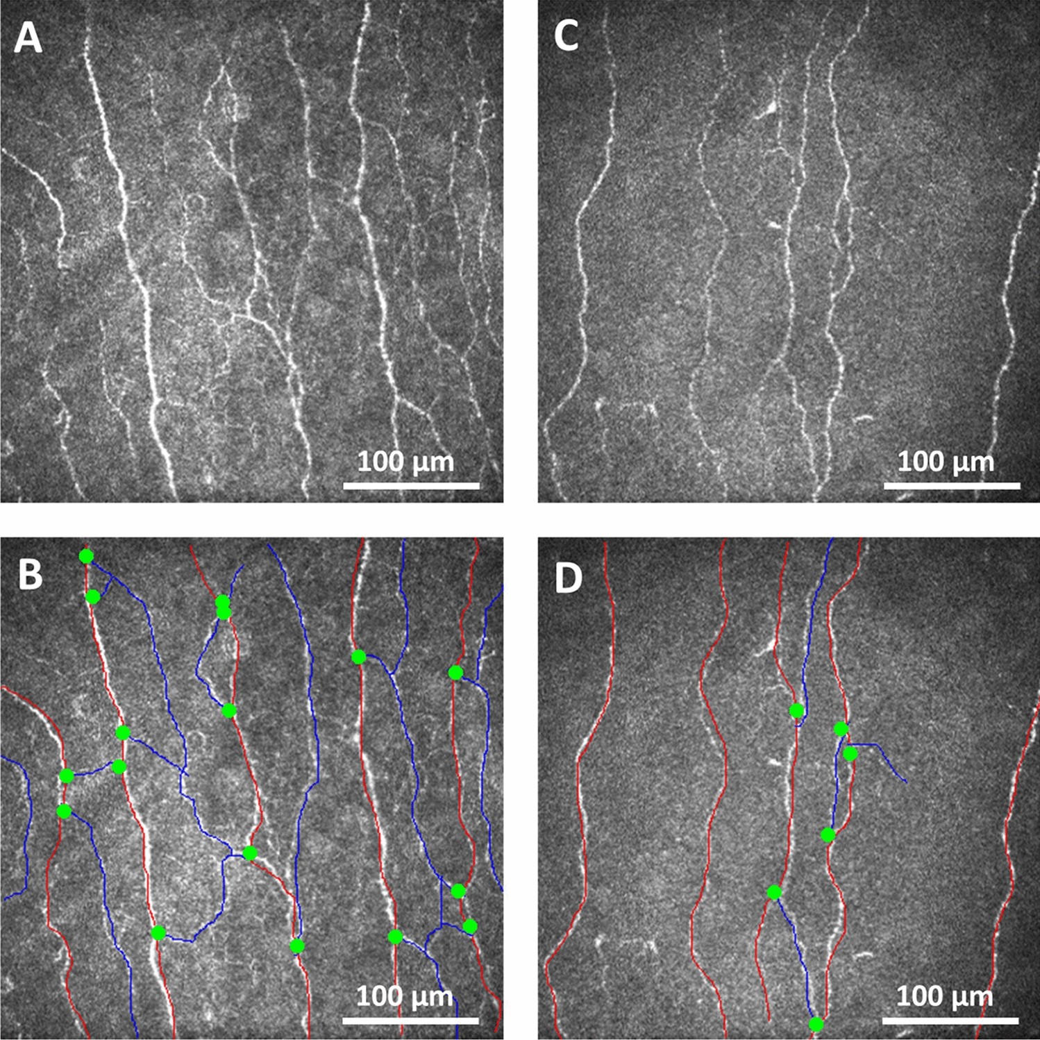
KIT - IAI - Abbildung des sub-basalen Nervenplexus der Kornea

Abnormal corneal nerve morphology and brain volume in patients with  schizophrenia | Scientific Reports
Abnormal corneal nerve morphology and brain volume in patients with schizophrenia | Scientific Reports